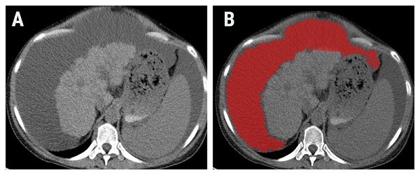
Ascites that occurs in oncological patients is often considered an indication of malignant invasion of the peritoneum. But in the absence of morphological changes associated with peritoneal carcinomatosis (PC), its cataloging based on the imaging aspect is almost impossible. It is desired that the cytological features of malignant ascites are also reflected in medical images, but are too subtle to be macroscopically assessed. The study published in BJBMS aimed to assess the ascites’ features on computed tomography (CT) images through a novel technique called texture analysis which quantifies the pixel intensity and distribution. This study included fifty-two patients with histologically proven benign (n=29) and malignant (n=23) intraperitoneal effusions.
The researchers used dedicated software (MaZda) that extracted specific texture parameters from the CT images. Their results suggest that, based on texture features, the differentiation between benign and malignant ascites was statistically significant. A texture parameter that reflects the local homogeneity of images held the highest discriminatory potential.
The researchers could not precisely identify the substrate that led to the textural differentiation between the two types of collections. Further studies are required to investigate whether these differences are determined by the malignant cellularity or other cytological, biochemical, or physical fluid properties.
If further validated, this method could represent a non-invasive diagnostic criterion for identifying early stages of peritoneal carcinomatosis, where ascites is the only sign. Also, it could spare the patient of the risks that come with invasive fluid sampling procedures.
Reference:
Editor: Edna Skopljak, MD
Leave a Reply