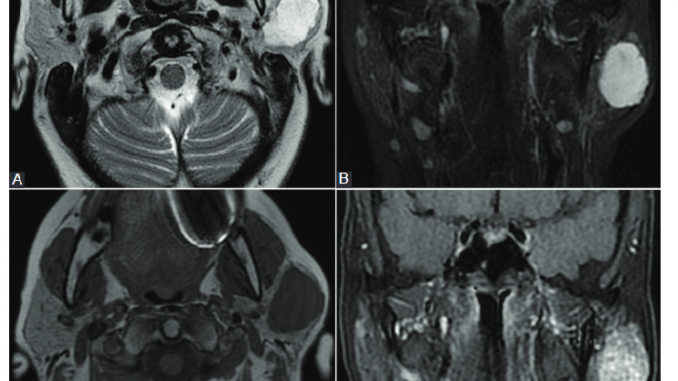
An accurate preoperative diagnosis of parotid tumors is essential for selection and planning of the surgical treatment, used in the most cases of parotid tumors. Many previous studies have shown that the modern cross-sectional imaging and cytologic investigations can support the preoperative differential diagnosis of parotid tumors.
The group of researchers from University of Medicine and Pharmacy and Emergency County Hospital, Cluj-Napoca aimed to achieve a comprehensive and updated review of modern imaging and cytologic investigations used in parotid tumors diagnosis, based on the latest literature data. Their paper, published in journal BJBMS was designed to serve as a guide for clinicians in selection of different cross-sectional imaging and cytologic investigations, in order to achieve an accurate preoperative differential diagnosis of parotid tumors.
After reviewing most of the articles published in the literature on this topic, the researchers concluded that magnetic resonance imaging (MRI) with its dynamic and advanced sequences represents the first-line imaging investigation used in diagnosis of parotid tumors. Computed tomography (CT) and positron emission tomography – computed tomography (PET–CT) have limited indications in differentiating parotid tumors. Fine needle aspiration biopsy and core needle biopsy can contribute with satisfactory results to the cytological diagnosis of parotid tumors.
Following this literature review, the researchers have drawn a series of conclusions with clinical applicability, as follows: dynamic MRI provides the best accuracy for preoperative differential diagnosis of parotid tumors, CT allows the best evaluation of bone invasion, being useful when MRI cannot be performed, and PET-CT is valuable in cancer patients follow-up. Furthermore, the dual, cytological and imaging approach is the safest method for an accurate differential diagnosis of parotid tumors.
Reference:
Editor: Edna Skopljak, MD
Leave a Reply