")
Dementia: More Than Just Memory Loss
Dementia is a formidable challenge in the medical landscape.
It’s worth noting that dementia isn’t a one-size-fits-all condition. Its progression can be segmented into stages:
- Early Stage: Often mistaken for age-related forgetfulness. However, subtle changes in memory, mood, and communication skills might hint at the onset of dementia.
- Middle Stage: The symptoms become more pronounced, with noticeable forgetfulness, confusion, and difficulties in daily tasks.
- Late Stage: Cognitive functions are severely affected, with patients often unable to recognize loved ones or communicate effectively.
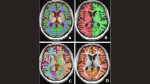
Types of Dementia and MRI Insights
Some of the most common types include:
- Alzheimer’s Disease: The most common form of dementia. MRI scans often reveal shrinkage in specific parts of the brain, primarily affecting memory.
- Vascular Dementia: Caused by reduced blood flow to the brain. MRI can spot the damaged areas, helping in diagnosis.
- Lewy Body Dementia: Characterized by abnormal protein deposits in the brain. MRI assists in distinguishing it from other types.
- Frontotemporal Dementia: Affects the frontal and temporal lobes. MRI highlights these specific areas of degeneration.
MRI’s Predictive Power
An Magnetic Resonance Imaging (MRI) machine consists of a large, circular tunnel with a table in the center. The patient lies on this table, and the machine generates a strong magnetic field around them. This magnetic field temporarily realigns hydrogen atoms in the body. Radio waves are then passed through the body, causing these aligned atoms to produce signals, which are then used to create images.
Common Uses of MRI
MRI has ability to produce detailed images without the use of harmful radiation, making them invaluable in diagnosing a range of conditions. These images can be in 2D or 3D and can be viewed from any direction, which provides a comprehensive view of the area being examined. Some of its common uses include:
Brain and Nervous System: MRI can detect brain tumors, traumatic injuries, developmental anomalies, multiple sclerosis, stroke, dementia, and infections. It offers a clear view of the brain and spinal cord and helps identify any abnormalities.
Bones and Joints: MRI can help in the diagnosis of conditions like arthritis, bone infections, bone tumors, and disc-related issues in the spine.
Breasts: An MRI of the breast (breast MRI) can be used alongside mammography to screen for breast cancer, especially in women who are considered high-risk.
Heart and Blood Vessels: MRI can assess the structure of the heart and aorta, looking for anomalies like aneurysms or tears. It can also assess the health and function of the heart chambers and valves.
Other Organs: MRI can visualize organs like the liver, kidneys, spleen, pancreas, and more. It can help detect tumors, cysts, and other abnormalities.
Tumors: MRI can be used to detect tumors throughout the body and is instrumental in planning their treatment.
Infections: MRI can help identify infections, especially in soft tissues.
One of the standout revelations from the Serbian team’s review is MRI’s potential to not just diagnose but predict dementia progression. By closely monitoring the brain’s changes over time, MRI can offer clues about the disease’s trajectory, enabling timely interventions.
The Global Impact of Dementia
With millions affected worldwide, the socio-economic implications are vast. Early diagnosis, facilitated by tools like MRI, can lead to better patient outcomes, reduced care costs, and more effective resource allocation in healthcare.
By looking at the brain’s structure and the pattern of any atrophy (wasting away of tissue), physicians can make informed decisions about the type and stage of dementia.
MRI scans can also rule out other conditions that might cause similar symptoms to dementia, such as brain tumors, subdural hematoma, or even stroke. Moreover, with the advent of advanced 3T MRI and techniques like arterial spin labeling, the visualization of minute changes in the brain due to early-stage dementia has become possible.
MRI not only aids in the early detection of the disease but also plays a role in monitoring its progression and assessing the effectiveness of treatments.
Practical Implications
The insights from the MRI scans aren’t just academic. They have real-world implications:
- Early Intervention: Recognizing the onset of dementia early can lead to timely treatments, potentially slowing the disease.
- Tailored Treatments: Understanding the specific type and stage of dementia ensures patients receive treatments best suited for their condition.
- Resource Allocation: For healthcare providers, knowing the prevalence and progression of dementia in their community can guide resource allocation and care planning.
Key Findings from the Review
- MRI’s Versatility: Beyond just diagnosis, MRI’s ability to track brain changes over time offers a deeper understanding of dementia’s progression.
- Consistency in Diagnosis: The introduction of semi-quantitative scales ensures MRI findings are consistent across the board.
- Potential for Early Detection: MRI’s ability to spot subtle brain changes, even in the early stages of dementia, is groundbreaking.
Conclusion: The Road Ahead
Dementia, with its profound impact on individuals and societies, demands our undivided attention. The review article by the researchers from Niš, Serbia, serves as a guideline to the community towards better understanding, diagnosis, and treatment. Through advanced MRI techniques, we’re not just witnessing the brain’s intricacies but are also equipped to respond, ensuring a brighter future for those affected by dementia.
The translation of the preceding English text in Serbian:
Demencija: Više od gubitka pamćenja
Demencija je ozbiljan izazov u medicinskom svetu.
Vredno je napomenuti da demencija nije bolest koja se manifestuje isto kod svih. Njen napredak može se podeliti na faze:
Rana faza: Često se povezuje sa zaboravnošću povezanom sa starenjem. Međutim, suptilne promene u pamćenju, raspoloženju i komunikacijskim veštinama mogu ukazivati na početak demencije.
Srednja faza: Simptomi postaju izraženiji, sa primetnom zaboravnošću, zbunjenošću i poteškoćama u svakodnevnim zadacima.
Kasna faza: Kognitivne funkcije su ozbiljno narušene, pacijenti često nisu u stanju da prepoznaju voljene osobe ili efikasno komuniciraju.
Tipovi demencije i uvidi iz MRI.
Neki od najčešćih tipova uključuju:
Alzhajmerova bolest: Najčešći oblik demencije. MRI skeniranja često otkrivaju smanjenje u specifičnim delovima mozga, prvenstveno utičući na pamćenje.
Vaskularna demencija: Uzrokovana smanjenim protokom krvi u mozak. MRI može detektovati oštećena područja, pomažući u dijagnozi.
Demencija s Lewy telima: Karakteriše je abnormalno taloženje proteina u mozgu. MRI pomaže u razlikovanju od drugih tipova.
Frontotemporalna demencija: Utice na frontalne i temporalne režnjeve. MRI ističe ova specifična područja degeneracije.
Prediktivna moć MRI
Aparat za magnetnu rezonancu (MRI) sastoji se od velikog, kružnog tunela sa stolom u centru. Pacijent leži na ovom stolu, a aparat generiše snažno magnetno polje oko njega. Ovo magnetno polje privremeno unosi vodikove atome u telo. Kroz telo se zatim šalju radio talasi, koji uzrokuju da ovi usklađeni atomi proizvode signale koji se zatim koriste za stvaranje slika.
Uobičajene primene MRI
MRI ima širok spektar primena u medicinskoj oblasti, zahvaljujući svojoj sposobnosti da proizvede detaljne slike bez upotrebe štetnog zračenja, čineći ih dragocenim za dijagnozu raznih stanja. Ove slike mogu biti u 2D ili 3D i mogu se posmatrati iz bilo kog pravca, što pruža sveobuhvatan pregled pregledanog područja. Neka od njihovih čestih upotreba uključuju:
Mozak i nervni sistem: MRI može detektovati tumore na mozgu, traumatske povrede, razvojne anomalije, multiplu sklerozu, moždani udar, demenciju i infekcije. Pruža jasan pogled na mozak i kičmenu moždinu i pomaže u identifikaciji bilo kakvih abnormalnosti.
Kosti i zglobovi: MRI može pomoći u dijagnozi stanja poput artritisa, infekcija kostiju, tumora na kostima i problema sa diskom u kičmi.
Grudi: MRI dojke može se koristiti uz mamografiju za skrining raka dojke, posebno kod žena koje se smatraju visokorizičnim.
Srce i krvni sudovi: MRI može proceniti strukturu srca i aorte, tražeći anomalije poput aneurizmi ili suza. Takođe može proceniti zdravlje i funkciju komora srca i ventila.
Drugi organi: MRI može vizualizovati organe poput jetre, bubrega, slezine, pankreasa i drugih. Može pomoći u detektovanju tumora, cista i drugih abnormalnosti.
Tumori: MRI se može koristiti za detekciju tumora u celom telu i ključan je za planiranje njihovog lečenja.
Infekcije: MRI može pomoći u identifikaciji infekcija, posebno u mekim tkivima.
Jedno od ključnih otkrića iz pregleda srpskog tima je potencijal MRI da ne samo dijagnostikuje, već i predviđa napredovanje demencije. Pažljivim praćenjem promena u mozgu tokom vremena, MRI može ponuditi tragove o putanji bolesti, omogućavajući pravovremene intervencije.
Globalni uticaj demencije
Demencija je globalni izazov. Sa milionima pogođenih širom sveta, socio-ekonomske implikacije su ogromne. Rana dijagnoza, olakšana alatima poput MRI, može dovesti do boljih ishoda za pacijente, smanjenih troškova nege i efikasnije raspodele resursa u zdravstvenoj zaštiti.
Gledajući strukturu mozga i obrazac bilo koje atrofije (propadanje tkiva), lekari mogu donositi informisane odluke o tipu i stadijumu demencije.
MRI skeniranja takođe mogu isključiti druge uslove koji bi mogli uzrokovati simptome slične demenciji, poput tumora na mozgu, subduralnog hematoma ili čak moždanog udara. Pored toga, sa dolaskom naprednog 3T MRI i tehnika poput oznake arterijskog spina, vizualizacija sitnih promena u mozgu zbog rane faze demencije postala je moguća.
MRI ne samo da pomaže u ranoj detekciji bolesti, već također igra ulogu u praćenju njenog napredovanja i proceni efikasnosti tretmana.
Detaljni uvidi koje pružaju MRI skeniranja su neprocenjivi, uvodeći eru informisane dijagnostike i planova lečenja.
Praktične implikacije
Uvidi iz MRI skeniranja nisu samo akademski. Oni imaju stvarne implikacije:
Rana intervencija: Prepoznavanje početka demencije rano može dovesti do pravovremenih tretmana, potencijalno usporavajući bolest.
Prilagođeni tretmani: Razumevanje specifičnog tipa i stadijuma demencije osigurava da pacijenti primaju tretmane koji su najbolje prilagođeni njihovom stanju.
Raspodela resursa: Za pružaoce zdravstvenih usluga, poznavanje prevalencije i napredovanja demencije u njihovoj zajednici može usmeriti raspodelu resursa i planiranje nege.
Ključni nalazi iz preglednog članka
Svestranost MRI: Osim samo dijagnoze, sposobnost MRI da prati promene u mozgu tokom vremena nudi dublje razumevanje napredovanja demencije.
Doslednost u dijagnozi: Uvođenje polukvantitativnih skala osigurava da su MRI nalazi dosledni širom sveta.
Potencijal za rano otkrivanje: Sposobnost MRI da otkrije suptilne promene u mozgu, čak i u ranoj fazi demencije, je revolucionarna.
Zaključak: Pogled napred
Demencija, sa svojim dubokim uticajem na pojedince i društva, zahteva našu nepodeljenu pažnju. Pregledni rad istraživača iz Niša, Srbija, služi kao smjernica ka boljem razumevanju, dijagnostikovanju i lečenju. Kroz napredne MRI tehnike, ne samo da svedočimo o složenostima mozga, već smo i opremljeni da reagujemo, obezbeđujući svetliju budućnost za one pogođene demencijom.
Reference: Živanović M, Aracki Trenkić A, Milošević V, Stojanov D, Mišić M, Radovanović M, Radovanović V. The role of magnetic resonance imaging in the diagnosis and prognosis of dementia. Biomol Biomed [Internet]. 2023Mar.16 [cited 2023Oct.7];23(2):209–224. Available from: https://www.bjbms.org/ojs/index.php/bjbms/article/view/8085
Editor: Ermina Vukalic
Leave a Reply