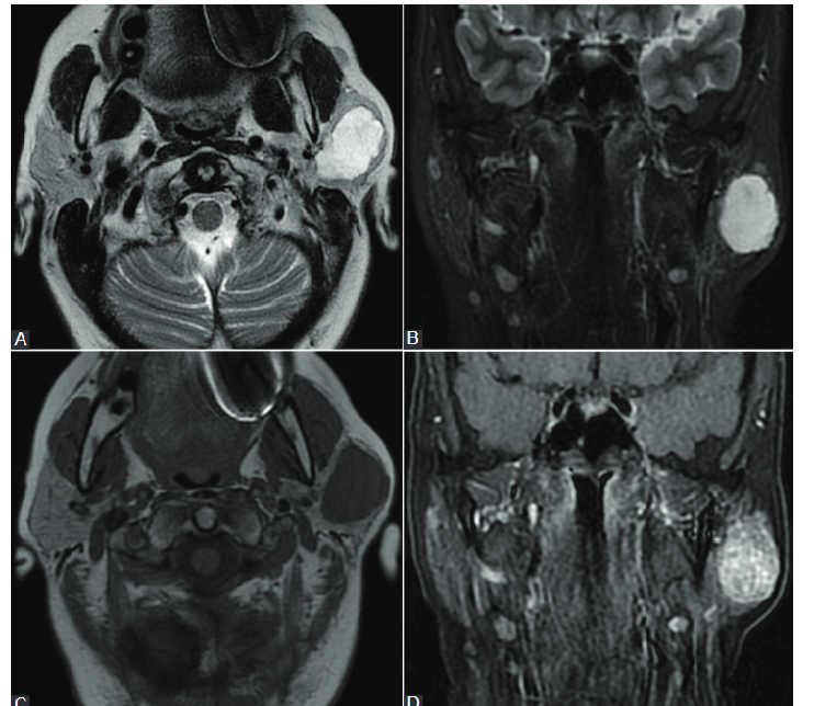
Conventional magnetic resonance imaging appearance in a 60-year-old patient with a left parotid gland pleomorphic adenoma. (A and B) T2-weighted image (WI) sequence and short tau inversion recover (STIR)-WI sequence revealing hyperintense signal. (C and D) T1-WI sequence without contrast and T1 contrast-enhanced sequences showing hypointense signal and homogeneous enhancement.
Untitled
Conventional magnetic resonance imaging appearance in a 60-year-old patient with a left parotid gland pleomorphic adenoma. (A and B) T2-weighted image (WI) sequence and short tau inversion recover (STIR)-WI sequence revealing hyperintense signal. (C and D) T1-WI sequence without contrast and T1 contrast-enhanced sequences showing hypointense signal and homogeneous enhancement.
Leave a Reply