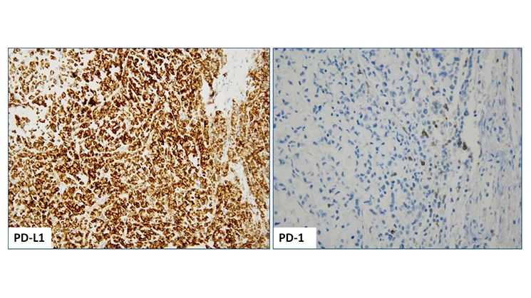
Positive PD-L1 (SP142 clone) IHC staining (2+, 85%) of a MSI-H colorectal tumor with concurrent BRAF V600E mutation; image taken at 20x magnification (Left image); Positive PD-1 (Cell Marque) IHC staining; image taken at 40x magnification (Right image).
Positive PD-L1 (SP142 clone) IHC staining (2+, 85%) of a MSI-H colorectal tumor with concurrent BRAF V600E mutation; image taken at 20x magnification (Left image); Positive PD-1 (Cell Marque) IHC staining; image taken at 40x magnification (Right image).
Positive PD-L1 (SP142 clone) IHC staining (2+, 85%) of a MSI-H colorectal tumor with concurrent BRAF V600E mutation; image taken at 20x magnification (Left image); Positive PD-1 (Cell Marque) IHC staining; image taken at 40x magnification (Right image).
Leave a Reply