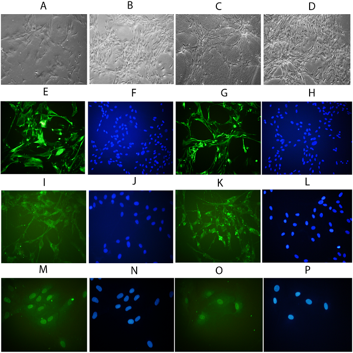
Photomicrographs of cultivated hRPE cells seeded on PG3 versus controls. Note the
epithelioid morphology of the control cells on days 3 (A) and 7 (B), and an elongated morphology
of the PG3-treated cells with dendritiform extensions on day 3 (C). The PG3 treated cells
demonstrate the epithelioid morphology on day 7 (D). Both control (E, I, M) and PG3-treated cells
(G, K, O) are stained positively (green) for the fluorescein isothiocyanate (FITC)-conjugated
cytokeratin 8/18 antibody (E & G), FITC-conjugated RPE65 antibody (I & K), and FITCconjugated
PAX6 antibody (M & O). Note DAPI-stained corresponding hRPE cells nuclei (blue)
in images F, J, and N for controls, and in images H, L, and P for PG3-treated cells. (Overall
magnification in all images × 200).
Photomicrographs of cultivated human retinal epithelial cells seeded on platelet gel versus controls.
Photomicrographs of cultivated human retinal epithelial cells seeded on platelet gel versus controls.
Leave a Reply