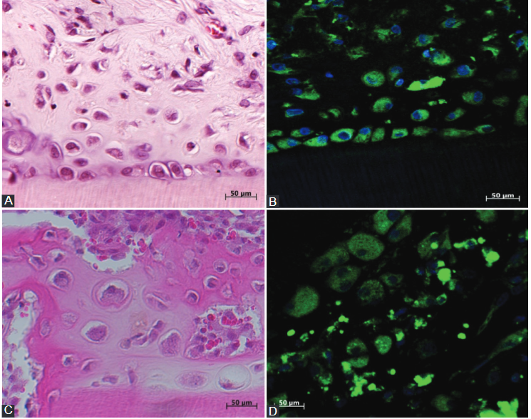
(A) Hematoxylin and eosin (H and E) staining of osteodentin-like hard tissue formed by dental pulp cells (DPCs) in the collagen gel after 21 days in the experimental group; (B) newly formed osteodentin inside demineralized dentin tubules. Osteoblast/odontoblast-like cells are showing positive immunofluorescence staining for green fluorescent protein in the experi-mental group; (C) H and E staining in the control group. DPCs formed a bone matrix-like hard tissue inside the walls of demineral-ized dentin tubules. H and E staining in the control group; (D) positive immunofluorescence staining of the osteoblasts inside the bone newly formed by DPCs without a collagen gel in the dentin tubules from the control group.
Untitled
(A) Hematoxylin and eosin (H and E) staining of osteodentin-like hard tissue formed by dental pulp cells (DPCs) in the collagen gel after 21 days in the experimental group; (B) newly formed osteodentin inside demineralized dentin tubules. Osteoblast/odontoblast-like cells are showing positive immunofluorescence staining for green fluorescent protein in the experi-mental group; (C) H and E staining in the control group. DPCs formed a bone matrix-like hard tissue inside the walls of demineral-ized dentin tubules. H and E staining in the control group; (D) positive immunofluorescence staining of the osteoblasts inside the bone newly formed by DPCs without a collagen gel in the dentin tubules from the control group.
Leave a Reply