
Anterior cervical discectomy and fusion represents one of the most common spinal surgeries. It is frequently used for the treatment of degenerative diseases of the cervical spine.
To our knowledge, there are no previously published peer-reviewed video cases of 3-level anterior cervical discectomy and fusion with detailed descriptions of the intraoperative microsurgical anatomy.
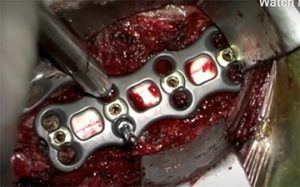
In the video article published in BJBMS, the authors present the case of a 33-year-old male who presented with bilateral myelopathy of the upper and lower extremities. The group performed a successful 3-level microsurgical anterior cervical discectomy and fusion from C4 to C7 vertebra. The patient was discharged home on the first postoperative day. His pain, numbness, and tingling resolved, as well as his myelopathy.
This case was submitted to demonstrate microsurgical anatomy, illustrate the operative technique used by the neurosurgeons in Semmes Murphey Neurologic and Spine Institute (Memphis, Tennessee, United States), which includes certain technical tips, and add to the literature.
We believe that this surgical video will aid other aspiring spinal neurosurgeons by providing a visual example of the 3-level ACDF procedure.
Reference:
Editor: Edna Skopljak
Leave a Reply